An Italian team of scholars has discovered an important new technique for larcheology
An all-Italian team has developed an innovative method that will revolutionize the field ofarchaeology. Scholars have used it with surprising results on archaeological bones, making the invisible visible. These important results, published in the journal Communications Chemistry of the Nature group, stem from extensive research work, by an Italian group, coordinated by Professor Sahra Talamo, in which experts in the field of analytical chemistry from the University ofBologna and theUniversity of Genoa collaborated.
This is a new technique for analyzing archaeological bones that for the first time allows for the quantification and high-resolution mapping of the presence of collagen, a protein that is essential for making radiocarbon dates and obtaining new information onhuman evolution.
“This innovation will allow significant progress in the study of human evolution,” explains Sahra Talamo, coauthor of the study and director of the BRAVHO Radiocarbon Dating Laboratory at the University of Bologna. “In fact, we will be able to analyze the valuable bone specimens and obtain precise dates while minimizing the amount of material taken.”
The bones of ancient hominids and bone jewelry found at archaeological sites are extremely valuable assets that should be carefully preserved and protected. Indeed, from these finds it is possible to obtain much information about the lives of ancient human populations: what they ate, their reproductive habits, their diseases, their movements and migrations. The possibility of obtaining this information, however, is linked to the amount of collagen present in bone artifacts.
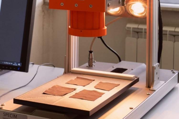
To combine the need to preserve the integrity of the artifacts as much as possible with the need to perform radiocarbon analyses, scholars, using a near-infrared associated camera, will be able to detect the average collagen content in the observed samples. “In this study, we have used imaging technology to quantify the presence of collagen in bone samples in a non-destructive way: it is thus possible to select the most suitable samples for radiocarbon dating analysis,” said Cristina Malegori, a researcher at the University of Genoa and first author of the paper. “Hyperspectral imaging in the near-infrared region was used to detect the distribution of collagen in ancient bones: a model that provides a chemical mapping of collagen content by quantifying its presence in each individual pixel.”
Sampling human foss ils and bone artif acts for radiocarbon dating is a destructive process, and human fossils or bone artifacts are increasingly rare and valuable as time goes on. Added to this is the fact that many of the archaeological bones are too small or too valuable to sample. It is therefore essential to be able to obtain preliminary, non-destructive information about the distribution of collagen in the artifacts to be analyzed. And the new technique fully meets this need, because it allows information to be obtained on both the location and content of collagen still present in a bone sample.
“The near-infrared hyperspectral imaging technique we used is a line-scanning system that acquires chemical images in which, for each pixel, a full spectrum in the near-infrared spectral range is recorded,” explained Giorgia Sciutto, professor at the University of Bologna and coauthor of the study. “The analysis system does not damage the specimen and allows reliable results to be obtained in a matter of minutes: thus, several specimens can be quickly analyzed to find suitable ones, avoiding the destruction of valuable material and greatly reducing time and costs.”
Scholars predict that this technique will also make it possible to perform radiocarbon dating at many archaeological sites where until now it had not been possible to analyze samples that had come to light due to their poor preservation.
“This new technique allows not only to select the best samples, but also to choose the sampling point in those selected according to the amount of collagen expected,” added Paolo Olivieri, professor at the University of Genoa and co-author of the study. “In this way it is possible to drastically reduce the number of samples used for radiocarbon analysis and, within the bone, it helps to avoid the selection of areas that might have an insufficient amount of collagen for dating.”
In fact, the survey can be performed either in small, localized areas or over the entire surface of the sample, thus producing a larger and much more meaningful amount of data.
“The potential of the proposed method lies in the type and amount of information the predictive model provides, answering two fundamental and complementary questions for the characterization of collagen in bone: how much and where,” confirms Cristina Malegori. “Knowing the amount of collagen concentrated in a precise area of the bone allows us to cut only this portion,” Professor Talamo concludes. “Also, when collagen prediction shows that the bone is poorly preserved, we can decide to perform targeted pretreatment to minimize collagen loss during extraction.”
The study was published in the journal Communications Chemistry under the title Near-infrared hyperspectral imaging to map collagen content in prehistoric bones for radiocarbon dating. The authors are Giorgia Sciutto, Silvia Prati, Lucrezia Gatti, Emilio Catelli, Silvia Cercatillo, Dragana PaleÄek, Rocco Mazzeo, Sahra Talamo (University of Bologna, Department of Chemistry “Giacomo Ciamician”), Stefano Benazzi (University of Bologna, Department of Cultural Heritage), Cristina Malegori, Paolo Oliveri (University of Genoa, Department of Pharmacy).
 |
| An Italian team of scholars has discovered an important new technique for larcheology |
Warning: the translation into English of the original Italian article was created using automatic tools. We undertake to review all articles, but we do not guarantee the total absence of inaccuracies in the translation due to the program. You can find the original by clicking on the ITA button. If you find any mistake,please contact us.


